Cervical Spine
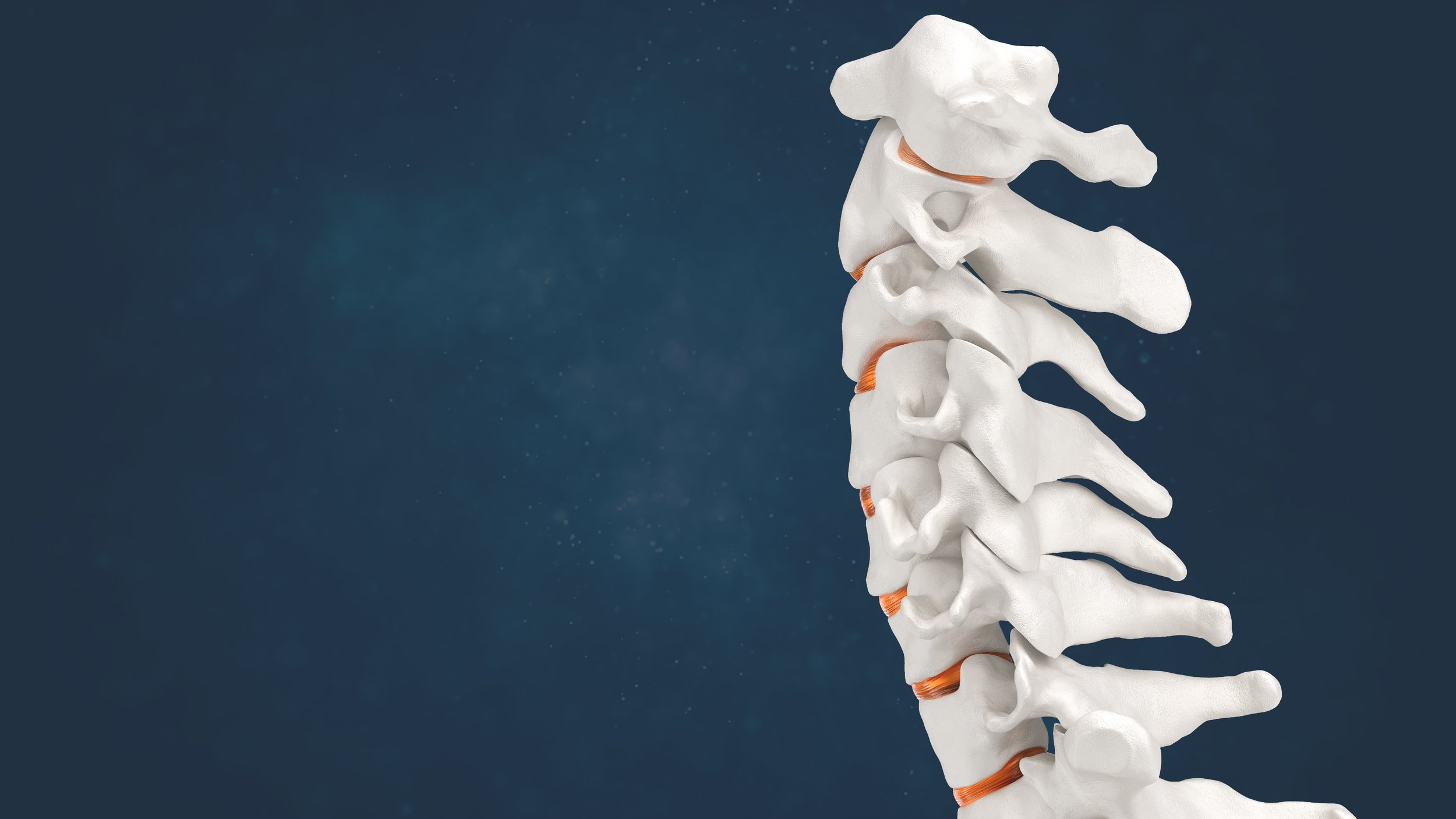
Anatomy of the Cervical Spine
C1-C7

About Cervical Spinal Degeneration
In the normal spine, the discs, located between the 7 neck bones (vertebrae) function as a shock absorber spacer, and for movement. Age and genetics can cause to damage to discs in the neck, causing the vertebrae to “pinch” the nerves exiting from behind the disc. In this case, the discs are degenerative, and the nerves are being impinged upon. The spinal cord itself may also be compressed. The compression narrows the pathway through which the nerves travel, and is called spinal stenosis. This can cause pain that can radiate into the neck, back, and arms and/or cause weakness and numbness.

Treatment
Anterior Cervical Disectomy and Fusion (ACDF)
The cervical spine includes the 7 bones and associated soft tissues of the neck. It is the most mobile portion of the spine, which makes it vulnerable to degenerative changes and injury. Non-surgical treatments are always used prior to performing surgery, unless the patient has sustained a life-threatening injury to the spine. These treatment measures include medication and physical therapy.
In the case of degeneration, most surgeries are performed from the front. The degenerated discs are removed and the spaces around the nerve structures are visualized and “cleaned” out. The disc is then replaced with a cage packed with crushed bone to re-establish the space available for the nerves and spinal cord that are located behind the disc. The bone, called a graft, will grow in and around the cage, connecting one vertebra to the next. This bony growth spanning the segment is called a spinal fusion. A titanium plate is commonly placed vertically along the front of the spine to buttress the cages.
For more specific questions please check out our Cervical Patient Brochure